Loading...

Generating PDF ...

  1. Endogenous Fields & Mind › 
  2. Endogenous Electromagnetic Fields › 
  3. Electromagnetism & Cancer
zoom-in section zoom-out section

Electromagnetism & Cancer
The break of coherence of endogenous electromagnetic fields is the key precursor of cancer

Pablo Andueza Munduate

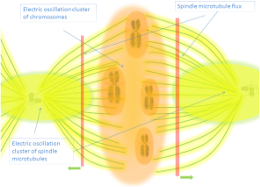
That endogenous electromagnetic (EM) fields are supporting life and its proper functioning can be viewed in how cancer cells generate variations of those internal EM fields that reveal their pathological state. Clusters of centrioles that vibrate longitudinally and coherent electric polar vibrations, dependents on water ordering in the cell, are some of the sources of these specific electromagnetic waves. ...

Investigations are bringing the view that cancer is a direct consequence of mitochondrial dysfunction, disturbed microtubular polar oscillations and the original coherent electromagnetic fields [1] (that include also magnetic fields generated by ionic transport at the membranes as we will see) that is extended and measurable at tissue scale also [2], in general it can be said that the cancer transformation pathway includes a link with altered coherent electric vibrations, and is believed that the individual independent activity begin when the frequency spectrum is rebuilt and shifted along with disturbances in the spacial pattern of the field [3].

Cancer cells can be classified depending on how they intake energy, viewing what processes are behind that energy generation that are altered respect to normal cells (altered energy transformation processes in cancer cells is a central phenomenon [4]). Two main branches can be classify here; those that obtain even more than 50 % of their ATP by metabolizing glucose directly to lactic acid even in the presence of oxygen (Warburg effect cancers, differentiated cancers) and those who are fueled by the supply of energy-rich metabolites such as lactate, pyruvate, glutamine and ketone BHB (beta-hydroxybutyrate) from the associated fibroblasts (reverse Warburg effect cancers, undifferentiated cancers). As is described in [5] in both cases the distorted energy supply alters the cell’s endogenous electromagnetic field transforming a normal cell into cancerous cell:

" In a Warburg effect cancer cell, the ordered water layer is reorganized and the water layer releases electrons, efficiently damping electromagnetic activity. The electromagnetic organization forces are weak, and coherence is disturbed. In the reverse Warburg effect cancers, the mitochondrial dysfunction and damped electromagnetic field occur in associated fibroblasts transporting energy-rich metabolites to the cancer cell whose electromagnetic field exhibits high energy with fluctuating power level and low coherence."

A variety of external agents, as is experimentally evidenced, can induce cancer, but their substrate, the physical reason of why can promote the disruption is in all the same [6]:

" Cancer can be initiated in a cell or a fibroblast by the short-circuiting of the cellular electromagnetic field by various fibers, parasitic energy consumption, virus infections, and mitochondrial defects, leading to a damped cellular electromagnetic field. Except short-circuiting (e.g., by asbestos fibers), the central process is mitochondrial dysfunction in cancer cells (the Warburg effect) or in fibroblasts associated with a cancer cell (the reverse Warburg effect), critically lowered respiration, reversed polarity of the ordered water layers around mitochondria emitting electrons, and damped electromagnetic activity of the affected cells."

It must be taken into consideration also the important role of water and its relation to EM field (for example resonant frequencies of microtubules in the frequency range of 10–30 MHz and 100–200 MHz are now discovered, and water inside microtubules is important as, for example, resonant peaks are not observed after release of water from the microtubule cavity) existing the viewpoint that cancer cells are progressed when cell’s interfacial water's normal state, that include the regular formation of water coherent domains (CD) (collections of liquid water molecules oscillating in tune with a self-trapped electromagnetic field at some defined frequency and that have a dedicated section on this site [7]), is disrupted as described in [8]:

" Both gene structure and protein structure, according to our thesis, are slaved to the biophysical status of interfacial water; hence, biomacromolecular structures react to supramolecular events. The proposed function of nanomolecular clusters of coherent water in water CDs is discussed. The hypothesis is presented that cationic Al, for example, effectively “short-circuits” the coherent nano-engines of our biomembranes, dramatically disrupting the delicately-balanced structural entropy consumption, necessary for charge separation, and transmission of both energy and information throughout the body. Concomitant increase in interfacial water stress and softening of tissues, with associated disruption of the cytoskeleton, has now been documented by multiple spectroscopic modalities."

It is know that some viruses are also linked to the generation of certain kinds of cancer but this can be explained as a result of parasitic virus energy consumption leading to randomized genome chemical bonding [9]:

" The ribonucleic acid (RNA) of the LDH virus parasitizing on energy reduces the ratio of coherent/random oscillations. Decreased effect of coherent cellular electromagnetic field on bonding electrons in biological macromolecules leads to elevating probability of random genome reactions."

On the other side, cancer cell’s external electromagnetic fields are enhanced to respect normal fields in a wide frequency spectrum, we have the electric fields generated by centrioles that in cancer cells are supernumerary and are clustered together causing the enhanced electromagnetic fields associated with cancerous tissue that in turn could attract blood vessels which then expand and also become malignant [4].

Moreover enhanced electric field can be detected also at tissue scales with electric currents of 0–10 μA/cm2 around the tumor [2], that are comparable to electric fields generated around wounds, with the difference that in tumors the direction and magnitude of these currents are inhomogeneous which may be due to the heterogeneity of local tumor tissue [2]. These fields can be responsible of the tumor’s unordered propagation dynamics [10]:

" The above data indicates that the “decision” of a certain part of the body to develop a tumor depends on the bioelectric state of remote regions. … It is important to mention that the same effect was obtained using any method of depolarization of Vmem (by modulating chlorine, sodium, potassium, or hydrogen channels). This in turn indicates the primary role of a purely physiological perturbation – disturbance in Vmem in the appearance of a metastatic phenotype, and not in case of any specific gene product or ionic disturbances."

Which has a very reasonable logic because electric fields are pivotal guiding cues for morphogenesis in general [11].

And not only electric fields but detectable magnetic fields are also shown to be altered, for example, in an interesting experiment where magnetic fields in the order of 1 to 100 pT are detected from the cellular ion transport through the cell membrane [2] being found that ion dynamics, and consequent magnetic fields, are different on cancer and non-cancer cells:

" It is interesting to observe that the nondifferentiated H9c2(2-1) rat cardiac myoblasts and the differentiated myocytes showed a significant difference in their magnetic signatures. The characteristic frequency of 27.8 Hz for the myoblasts was found to be identical to that of HeLa cells, which is a cancer cell line. The differentiated cell lines showed a cluster of higher characteristic frequencies of around 220 Hz. It is speculated that each of the three frequencies in the cluster is attributable to a specific ionic flux. One could then infer that the ion channel-gating dynamics of nondifferentiated cell lines are nondistinct, which is a key factor affecting cellular homeostasis."

In another frequency spectrum there is also an increment in biophotonic emissions in cancer cells as it's described, for example, in this investigation [12]:

" Only 1% of malignant cells in a normal aggregate, representative of the early stages of cancer development, resulted in conspicuously increased numbers of photon emissions and spectral power spectra that often reflect a total malignant cell population. This combination of photon flux density and spectral power profiles may be a potentially useful (nanotechnology) tool to detect the minute changes in cell activities relevant to oncology."

Posterior investigations delves into this discovery [13] (there is a subsection [14] dedicated to biophotonic characteristic emissions from cancer cells):

" Using wavelength-exclusion filters, we demonstrate that ratios between infrared and ultraviolet photon emissions differentiate cancer and non-cancer cell types. Further, we identified photon sources associated with three filters (420-nm, 620-nm., and 950-nm) which classified cancer and non-cancer cell types. The temporal increases in biophoton emission within these wavelength bandwidths is shown to be coupled with intrisitic biomolecular events using Cosic's resonant recognition model."

And this is important because it is possible that cancerous cells can recruit other cells through biophotonic information also; as is pointed out in [15] one thing is the amount of energy necessary to maintain the molecular machinery, that uses glucose metabolism-based chemical processes, and other thing the possible non-local determinants of the information that initiates or terminates these processes, that can be in form of biophotons (in the paper compare them with the small effort of turning the key in the ignition on a combustion motor car).

So as in other sections of this web we are speaking about various layers of electromagnetic fields whose distortion, in this case, can cause the cancer initiation and propagation.

References:

1. Pokorný, J., Pokorný, J., & Kobilková, J. (2013). Postulates on electromagnetic activity in biological systems and cancer. Integrative Biology, 5(12), 1439-1446.

2. Zhu, K., Hum, N. R., Reid, B., Sun, Q., Loots, G. G., & Zhao, M. (2020). electric fields at Breast cancer and cancer cell collective Galvanotaxis. Scientific Reports, 10(1), 1-11.

3. Pokorný, J., Pokorný, J., Kobilková, J., Jandová, A., Vrba, J., & Vrba Jr, J. (2014). Cancer—Pathological breakdown of coherent energy states. Biophysical Reviews and Letters, 9(01), 115-133.

4. Huston, R. L. (2016). A Review of Electromagnetic Activity in Cellular Mechanics. Advances in Bioscience and Biotechnology, 7(9), 360-371.

5. Pokorný, J., Pokorný, J., & Borodavka, F. (2017). Warburg effect—damping of electromagnetic oscillations. Electromagnetic Biology and Medicine, 36(3), 270-278.

6. Pokorný, J., Pokorný, J., Kobilková, J., Jandová, A., & Holaj, R. (2020). Cancer Development and Damped Electromagnetic Activity. Applied Sciences, 10(5), 1826.

7. MMIND › Endogenous Fields & Mind › Water & Electromagnetic Fields › Electromagnetism & Water - Coherence Domains

8. Davidson, R. M., Lauritzen, A., & Seneff, S. (2013). Biological water dynamics and entropy: a biophysical origin of cancer and other diseases. Entropy, 15(9), 3822-3876.

9. Pokorný, J., Pokorný, J., Jandová, A., Kobilková, J., Vrba, J., & Vrba Jr, J. (2016). Energy parasites trigger oncogene mutation. International journal of radiation biology, 92(10), 577-582.

10. Draguţa, I., Mustea, A., Popescu, C., Iurcu, C., & Palade, V. (2019). Disturbance of bioelectric transmission in carcinogenesis. Moldovan Medical Journal, 62(2), 33-37.

11. EMMIND › Endogenous Fields & Mind › Endogenous Electromagnetic Fields › Electromagnetism & Morphogenesis

12. Karbowski, L. M., Murugan, N. J., Dotta, B. T., & Persinger, M. A. (2015). Only 1% Melanoma Proportion in Non-Malignant Cells Exacerbates Photon Emissions: Implications for Tumor Growth and Metastases. Int J Cancer Res Mol Mech, 1(2).

13. Murugan, N. J., Rouleau, N., Karbowski, L. M., & Persinger, M. A. (2018). Biophotonic markers of malignancy: Discriminating cancers using wavelength-specific biophotons. Biochemistry and biophysics reports, 13, 7-11.

14. EMMIND › Endogenous Fields & Mind › Biophotons › Biophotons - Various › Biophotons differently emitted by cancer cells

15. Dotta, B., Buckner, C., & Lafrenie, R. (2016). Photon Emissions as differ‐ential indicators for different components of protein kinase A (PKA) in transfected murine melanoma cells. Arch can Res, 4(3), 10-21767.

Very related sections:

expand upper introductory text Generate PDF ⇊ Paginate ≣

text updated: 26/06/2020
tables updated: 22/10/2024

Endogenous Fields & Mind
EM & Cancer

Endogenous Electromagnetism & Cancer

(F) Full or (A) Abstract

Available Formats

Title

Commentary

Publication Year (and Number of Pages)

Author(s)
SEE ALSO THIS (subsection on ELF applied to cancer cells)
OR THIS (subsection on biophotons differently emitted by cancer cells).
Favailable in PDF and HTMLBiophysical control of plasticity and patterning in regeneration and cancerCommentary icon2023-(23)Nirosha J. Murugan, Solsa Cariba, Sawith Abeygunawardena, Nicolas Rouleau, Samantha L. Payne
Favailable in PDF and HTMLViolation of molecular structure of intracellular water as a possible cause of carcinogenesis and its suppression by microwave radiation (hypothesis)Commentary icon2023-(6)Vitali Kalantaryan, Radik Martirosyan, Yuri Babayan, Voldemar Petrosyan
Favailable in PDFPhysical aspects of chemical carcinogenesisCommentary icon2023-(23)A. P. Batyanov
Favailable in PDF and HTMLUHF-Dielectrophoresis Microfluidic Lab-on-a-Chip to Detect the Transformation Potential of Extracellular Vesicles Derived From Cancer Stem CellsCommentary icon2022-(21)Élodie Barthout, Élisa Lambert, Céline Hervieu, Sofiane Saada, Rémi Manczak, Julie Pannequin, Arnaud Pothier, Claire Dalmay, Fabrice Lalloué, Muriel Mathonnet, Barbara Bessette
Favailable in PDF and HTMLA computational model of organism development and carcinogenesis resulting from cells’ bioelectric properties and communicationCommentary icon2022-(14)Joao Carvalho
Favailable in PDFThe Understanding of Enigmas Related to Cancer: Cancer Origin and Progression- A ReviewCommentary icon2021-(9)M. T. Hasan
Favailable in PDF and HTMLA bioelectric model of carcinogenesis, including propagation of cell membrane depolarization and reversal therapiesCommentary icon2021-(11)Joao Carvalho
Aavailable in HTMLBioelectrical approaches to cancer as a problem of the scaling of the cellular selfNo comments yet icon2021-(1)Michael Levin
Favailable in PDF and HTMLElectric Fields at Breast Cancer and Cancer Cell Collective GalvanotaxisCommentary icon2020-(11)Kan Zhu, Nicholas R. Hum, Brian Reid, Qin Sun, Gabriela G. Loots, Min Zhao
Favailable in PDF and HTMLCancer Development and Damped Electromagnetic ActivityCommentary icon2020-(17)Jiří Pokorný, Jan Pokorný, Jitka Kobilková, Anna Jandová, Robert Holaj
Favailable in PDF and HTMLMeasuring Cellular Ion Transport by MagnetoencephalographyCommentary icon2020-(8)Sudhir Kumar Sharma, Sauparnika Vijay, Sangram Gore, Timothy M. Dore, Ramesh Jagannathan
Favailable in PDF and HTMLBioelectric Control of Metastasis in Solid TumorsCommentary icon2019-(17)Samantha L. Payne, Michael Levin, Madeleine J. Oudin
Favailable in PDFDisturbance of bioelectric transmission in carcinogenesisCommentary icon2019-(5)Ilarion Draguta, Anatolie Mustea, Constantin Popescu, Cornel Iurcu, Valeriu Palade
Aavailable in HTMLStem Cell Differentiation Stage Factors and their Role in Triggering Symmetry Breaking Processes during Cancer Development: A Quantum Field Theory Model for Reprogramming Cancer Cells to Healthy PhenotypesNo comments yet icon2019-(1)P.M. Biava, F. Burigana, R. Germano, P. Kurian, C. Verzegnassi, G. Vitiello
Favailable in PDFCatalase intrinsic emissions of electromagnetic fields as probable cause in cancerogenesis from consumption of red and processed meatNo comments yet icon2018-(8)Abraham A. Embi
Favailable in PDF and HTMLWarburg effect—damping of electromagnetic oscillationsCommentary icon2017-(9)Jiří Pokorný, Jan Pokorný, Fedir Borodavk
Favailable in PDFCancer is promoted by cellular states of electromagnetic decoherence and can be corrected by exposure to coherent non-ionizing electromagnetic fieldsNo comments yet icon2017-(45)D.K.F. Meijer, J.H. Geesink
Favailable in PDF and HTMLEndogenous electromagnetic forces emissions during cell respiration as additional factor in cancer originNo comments yet icon2016-(3)Abraham A. Embi
Favailable in PDF and HTMLA Review of Electromagnetic Activity in Cellular MechanicsCommentary icon2016-(12)Ronald L. Huston
Favailable in PDF and HTMLEnergy parasites trigger oncogene mutationCommentary icon2016-(7)Jiří Pokorný, Jan Pokorný, Anna Jandová, Jitka Kobilková, Jan Vrba, Jan Vrba Jr
Favailable in PDF and HTMLMitochondrial Dysfunction and Disturbed Coherence: Gate to CancerNo comments yet icon2015-(21)Jiří Pokorný, Jan Pokorný, Alberto Foletti, Jitka Kobilkova, Jan Vrba, Jan Vrba Jr
Favailable in PDF and HTMLUsing the Electromagnetics of Cancer’s Centrosome Clusters to Attract Therapeutic NanoparticlesCommentary icon2015-(10)Ronald L. Huston
Aavailable in HTMLOn Centrioles, Microtubules, and Cellular ElectromagnetismCommentary icon2014-(1)Ronald L. Huston
Favailable in PDFOncology and Biophysics: A Need for IntegrationNo comments yet icon2014-(6)Sarah S. Knox, Richard H.W. Funk
Favailable in PDFCancer - pathological breakdown of coherent energy statesNo comments yet icon2014-(19)Jiří Pokorný, Jan Pokorný, Jitka Kobilková, Anna Jandová, Jan Vrba, Jan Vrba Jr.
Favailable in PDF, HTML and EpubBiophysical Insights into Cancer Transformation and TreatmentCommentary icon2013-(11)Jiří Pokorný, Alberto Foletti, Jitka Kobilková, Anna Jandová, Jan Vrba, Jan Vrba Jr., Martina Nedbalová, Aleš Čoček, Andrea Danani, Jack A. Tuszyński
Favailable in PDF and HTMLPostulates on electromagnetic activity in biological systems and cancerCommentary icon2013-(8)Jiří Pokorný, Jan Pokorný, Jitka Kobilková
Favailable in PDF, HTML and EpubEndogenous Voltage Potentials and the Microenvironment: Bioelectric Signals that Reveal, Induce and Normalize CancerNo comments yet icon2013-(60)Brook Chernet, Michael Levin
Favailable in PDF and HTMLBiological Water Dynamics and Entropy: A Biophysical Origin of Cancer and Other DiseasesCommentary icon2013-(55)Robert M. Davidson, Ann Lauritzen, Stephanie Seneff
Favailable in PDFThe Role of Coherence in a Systems View of Cancer DevelopmentNo comments yet icon2012-(33)M. Plankar, E. Del Giudice, A. Tedeschi, I. Jerman
Favailable in PDF and HTMLCancer physics: diagnostics based on damped cellular elastoelectrical vibrations in microtubulesNo comments yet icon2011-(13)Jiří Pokorný, Clarbruno Vedruccio, Michal Cifra, Ondřej Kučera
Favailable in PDFEmbryonic Morphogenetic Field Induces Phenotypic Reversion in Cancer Cells. Review ArticleCommentary icon2011-(11)M. Bizzarri, A. Cucina, P. M. Biava, S. Proietti, F. D’Anselmi, S. Dinicola, A. Pasqualato, E. Lisi
Favailable in PDFA chronic decrease of heart rate variability can precede some cases of cancerCommentary icon1997-(9)Sv. Danev, S. Svetoslavov, E. Datzov

.

.

Go to top of the page